Sports Injuries
17.4K views | +0 today
Follow
Sports Injuries
Sports injuries occur when participating in sports or physical activities associated with a specific sport, most often as a result of an accident. Sprains and strains, knee injuries, Achilles tendonitis and fractures are several examples of frequent types of sport injuries. According to Dr. Alex Jimenez, excessive training or improper gear, among other factors, are common causes for sport injury. Through a collection of articles, Dr. Jimenez summarizes the various causes and effects of sports injuries on the athlete. For more information, please feel free to contact us at (915) 850-0900 or text to call Dr. Jimenez personally at (915) 540-8444. http://bit.ly/chiropractorSportsInjuries Book Appointment Today: https://bit.ly/Book-Online-Appointment
Your new post is loading...
Scooped by Dr. Alex Jimenez
Scoop.it!

Foot Injuries: Scientific Perspective | El Paso Back Clinic® • 915-850-0900

Foot Injuries: Scientific Perspective | El Paso Back Clinic® • 915-850-0900 | Sports Injuries | Scoop.it

Injury specialist, Dr. Alexander Jimenez reviews the middle and forefoot anatomy -- also discusses several common athletic injuries.


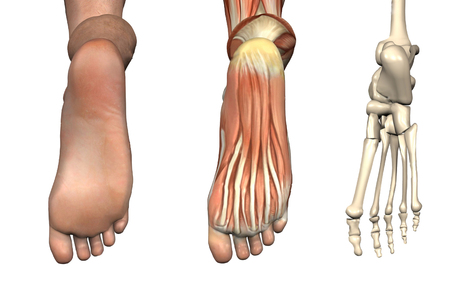
The foot consists of 26 interconnecting bones. The intricate anatomy of the foot makes it to be both a passive service structure and an active vehicle for force production (see Figure 1).


The mid foot begins at the transverse tarsal, or midtarsal, combined. The talus, navicular, calcaneus, and cuboid bones include this joint. The connections between these bones form an 'S'-shaped single joint line across the waist of the foot, with the navicular bone itself function as the structural link between the forefoot and hindfoot. The multiple articulations within the joint permit the forefoot to move independently in response to hindfoot movement. All five rays, comprised of the metatarsals and phalanges, work in concert to maximize foot stability.

Navicular Stress Fracture

A once obscure identification, navicular stress fractures, now account for 14% to 35 percent of all reported stress fractures(1). It's a particularly widespread investigation in runners, and athletes that execute volatile jumping, as in track and field sports. Suspect a navicular stress fracture in an athlete that complains of obscure mid foot pain but who can not remember actually hurting his foot. Palpation of the apex of the dorsal aspect of the navicular, called the 'N' place, causes pain in 81 percent of patients having a stress fracture(1).


The navicular lies at the crossroads of their longitudinal and transverse arches, and thus endures a unique utter stress in the middle third of the bone, where it's the most susceptible to fracture. The center third of the navicular is also the area with the least amount of vascularization. For this reason, fracture in this website resolves slowly. Treatment requires at least fourteen days in a non-weight posture throw(1).


One to 2 weeks following boot elimination, begin running and sport-specific training as tolerated, with weekly clinical evaluation of the navicular. If tenderness over the palpated navicular yields, refer for further immobilization or surgery. With conservative therapy, athletes typically return to game in five to six months, compared to three to four weeks for people who undergo surgical fixation(1).

Lisfranc Joint Injury

Named after Napoleon's field surgeon Jacques Lisfranc, who first observed this mid foot injury in men who had dropped from their horse in battle, this injury is due to forced plantar flexion at the joint. In today's sports, the mechanism of trauma is an axial load put on the elevated heel, usually from a fall or tackle by a different player (see Figure 2). Lisfranc injury is not uncommon in American football players (especially lineman in blocking stance, up on feet), football players, bicyclists, surfers, and equestrians. In sports which require feet in straps or clips, a Lisfranc injury may result in the entrapped forefoot in a fall.


An injury to the Lisfranc joints most commonly occurs medially, with a consequent separation of the first and second metatarsals, along with a dorsal displacement of the second metatarsal on the next cuneiform. Trainers with a Lisfranc injury normally present with significant pain, moderate to severe swelling, and bruising at the mid foot. Initial radiographic studies don't detect joint diastasis in up to a third of cases; therefore, weight bearing x-rays are justified if Lisfranc injury is suspected(2). An uncommon harm to the lateral Lisfranc joints happens as result of forced supination, and usually with no dislocation of the fracture or joints.


When just the ligaments of the joint are injured, care for the sprain conservatively. Begin weight bearing as tolerated but protect the joint by hammering the foot, using an orthotic shoe insert, or, in severe circumstances, immobilizing the joint using a walking fracture boot. Advance the athlete to cross- training activities, like swimming or biking, as soon as possible to keep fitness. Progress rehab to shut chain activities as tolerated. The athlete might need to utilize joint protection through taping, a shoe insert, or a stiff- soled shoe for up to 12 months(3). If the injury does not significantly improve after several weeks, ask repeated x-ray assessment in a weight bearing position.


Post-operatively, the athlete is generally non-weight posture for three to six weeks; however, encourage range of motion exercises with the feet, even while immobilized from the post-operative splint. Screw removal takes place between three to four months post-procedure, depending upon individual protocols. Rehabilitate with speed, strength, and endurance training as well as tolerated. Return to play just when the athlete illustrates closed-chain plantar flexion strength and single limb equilibrium appropriate for their sport, normally within four to six months.

Metatarsal Stress Fracture

During extended periods of training, muscles on the bottom of the foot fatigue and fail to protect the metatarsals from floor reaction forces. With no plantar muscles to dissipate this stress, the bones suffer recurrent micro-trauma that results in stress fractures.


Athletes complain of obscure forefoot pain that resolves with rest, but intensifies with continued instruction. The forefoot might be bloated and bruised. X-rays performed shortly after symptoms begin may not demonstrate a fracture; however, a bone scan or magnetic resonance image (MRI) often reveals the severe injury.


These fractures usually respond well to conservative therapy with weight bearing as tolerated but restricted activity for four to six weeks. Following six weeks, restart game training as tolerated and track for a return of symptoms. Using custom orthotics corrects biomechanical problems, such as flat feet, which could induce the athlete to repeated injury.

Dancer's Fracture

An avulsion fracture at the tuberosity at the base of the fifth metatarsal may be known as a 'dancer's fracture'. Within an avulsion fracture, the athlete complains of pain, bruising, and swelling in the base of the fifth metatarsal. This injury often accompanies a foot sprain.


Simple avulsion fractures respond well to conservative treatment of rest, ice, compression and protection. Permit weight bearing as tolerated at a splint or walking boot. Symptoms usually improve within six to eight months, at which time progressive return to sport training begins. Monitor the athlete clinically for any signs of mal-union of the fracture. Avulsions with displacement are treated surgically.

Jones Fracture

Acute fractures of the shaft of the fifth metatarsal are known as Jones fractures, called after Sir Robert Jones who first described this fracture in his own foot in 1902(4). Jones fractures happen if the heel is off of the floor and also a force is applied to the forefoot in the lateral direction, causing the ankle to reverse and roll cartilage. This region is minimally vascularized and are also the website of repetitive micro-trauma. This sort of fracture happens in amateurs, footballers, baseball and hockey players, who commonly pivot to a plantar flexed foot.


An athlete with a Jones fracture poses with pain, swelling, and bruising at the bottom of the fifth metatarsal. Weight bearing may be difficult. Fractures are categorized into three kinds. Type I is a serious fracture with clean margins and no displacement, and responds well to conservative therapy. Because of the decreased blood supply to that area of the bone, recovery takes at least six to eight months, and upward of 20 weeks to get complete calcification.


A fracture with delayed recovery is classified as a Type II. The fracture line is widened and X-ray reveals increased bone reabsorption at the margins of the fracture. A Form III is absolute non-union of the fracture. Types II and III may be because of repetitive micro trauma at the website prior to a serious event, or even the lack of vascularization which delays recovery from a serious injury. Post- surgical protocols differ, but most progress to partial weight bearing in a walking boot in one or two weeks. Athletes may start partial weight bearing activities, such as aquatic biking and training, at about fourteen days post-op and start working with shoe alterations or orthotics at six to eight weeks after surgery. Most return to sport by 12 weeks post-op.

Sesamoid Injury

The sesamoid bones are two oval-shaped bones inserted in the flexor hallucis brevis tendon, at the distal end of the first metatarsal. These bones function to lengthen the first metatarsal and assist with weight change and toe off through gait. As such, they suffer great stress in athletes that repeatedly 'push off' with the ankle in plantar flexion and the forefoot dorsiflexed, such as runners, dancers, football lineman, and golfers.


Of all sesamoid accidents, stress fracture happens 40 percent of the time, whilst sesamoiditis and acute fracture appear in just 30 percent and 10% of cases, respectively(5). As with other repetitive stress injuries, the onset of pain using a stress fracture could be insidious and vague. Passively bending the big toe can reproduce the pain. X-ray evaluation isn't always conclusive in such harms; therefore, a bone scan or MRI may be necessary for diagnosis.


Treatment consists of rest from the offending activity together with custom orthotics or a metatarsal pad to eliminate weight bearing directly to the bones. For athletes using sesamoiditis, an inflammatory condition caused by repeated stress to the bones, treat with rest, ice hockey and protection, and gradually initiate a return to action once they bear weight without pain. For those who have actual fracture, six months of non-weight bearing is necessary. If the fracture does not heal adequately following this time, consult with surgery. Athletes possibly return to sport within six months after surgery, which either partly or entirely eliminates the bone, or even fixes the fracture.

'Turf Toe'

Dancers, football lineman, rugby players, golfers, and wrestlers often assume a position of MTP joint dorsiflexion with ankle plantar flexion (see Figure 3). More and more flexible shoes, along with artificial turf surfaces, have been implicated in the greater incidence of this harm.


Participants present following an acute event with pain and tenderness over the joint, accompanied by swelling and bruising when the injury is categorized as Grades II (partial tear of the ligaments) or III. Conservative therapy with immobilization of the MTP joint through taping or bracing; protection with steel shoe inserts; progressive weight bearing; and restricted activity is the normal treatment course for all MTP harms. Initiate progressive sports instruction after three to six weeks as tolerated, and monitor for a recurrence of symptoms. Surgery is warranted only in the most extreme injuries with joint dislocation or harm to the sesamoid bones.

Morton's Neuroma

More common in women than men, Morton's neuroma is a nerve entrapment disease of the nerve between the toes. The nerve becomes thick and painful because of chronic friction together with the adjacent ligaments, for example happens when pivoting in tennis or dancing. Abnormal foot body is thought to contribute to the problem. Athletes whine of burning pain between the feet, at the metatarsal heads.


The pain can improve with rest and worsen with activity. A clinical diagnosis can be made with the lateral squeeze evaluation (see Figure 4).


Since not a true neuroma, but instead a result of entrapment or compression, sneakers with a large toe box generally improve symptoms. Conservative physiotherapy modalities are sometimes helpful. Therapeutic exercises may also help fortify the foot and reduce strain to the injured nerve. When all else fails, surgical decompression or resection of the nerve eliminates the pain, but results in a level of numbness to the area.

Conclusion

Injuries into the mid and forefoot result in a significant number of lost playing time in all sports. By some reports, sports- related injuries to the toes comprise 2% to 18 percent of all athletic injuries(6). While many of those mid and forefoot accidents occur needlessly, they do so in regions already exposed, due to either a decreased blood supply and also the biomechanics of the foot structure.


To minimize these injuries, use orthotics, taping, and therapeutic exercise to correct biomechanical and structural abnormalities inside the foot. Guide your athletes at proper shoe selection, eschewing whatever the shoe business rep could possibly be telling them is so popular. Look closely at functional deficits in gait and regions of relative weakness or strength imbalance. 1 weak gluteus medius muscle sends ripples down the biomechanical string that could leave a participant pivoting on a foot under more pressure than necessary.


References
1. Am Fam Physician. 2003 Jan 1;67(1):85-91.
2. BMJ. 2013 July;3,347:f4561.
3. Clin Sports Med. 1997 July;16(3):435-65.
4. Am Fam Physician. 1999 May 1;59(9):2516-22.
5. Curr Sports Med Rep. Sept/Oct 2011;10(5):249-55.
6. Sports Health. 2012 Nov;4(6):518-34.

Dr. Alex Jimenez's insight:

Injury specialist, Dr. Alexander Jimenez reviews the middle and forefoot anatomy -- also discusses several common athletic injuries. For Answers to any questions you may have please call Dr. Jimenez at 915-850-0900

No comment yet.
Scooped by Dr. Alex Jimenez
Scoop.it!

Foot Injuries: Lisfranc Joint (Part I) | El Paso Back Clinic® • 915-850-0900

Foot Injuries: Lisfranc Joint (Part I) | El Paso Back Clinic® • 915-850-0900 | Sports Injuries | Scoop.it

Chiropractor, Dr. Alexander Jimenez looks at injuries in the Lisfranc joint -- and how they may be treated.


Foot injuries are common in the athlete and they appear in many forms. These include sprains into the joints between the tarsals and metatarsals , chronic soft tissue injuries such as plantar fasciitis and stress fractures of the metatarsals and tarsal bones. Injuries to the midfoot and in particular the Lisfranc joint (or tarsometarsal joint) are a rare but potentially devastating injury to the sportsperson that require particular attention by the sports medicine practitioner.


Athletes were not involved by the type of the injury but involved personnel. Surgeon Jacques Lisfranc Saint Martin first described it who performed midfoot amputations on soldiers through the 'Lisfranc' joint after they'd dropped off their horses.


In present day scenarios, accidents to the joint are often high velocity injuries that cause severe deformity into the midfoot joints due to dislocations. These energy mechanics would be the result of a car/motor bike Collision, severe fall on the foot or a severe injury to the foot usually lasted in an industrial setting. From the athlete, injury severity is usually far less devastating and results from some form of crushing and/or twisting mechanism. But if it occurs it can have devastating consequences for the athlete.

Introduction

Although foot injuries in the athlete are reasonably common (16 percent of sports-related accidents involve the foot -- Garrick and Requa 1988), accidents to the Lisfranc joint are extremely rare. Myers et al (1994) discovered that Lisfranc injuries account for 4 percent of collegiate-level football injuries. However, the forms of the harm can't just be career-ending although season-ending for the athlete.


It is more typical in sports like American Football (using a high percentage of Lisfrancs happening in offensive linemen -- Myers et al 1994) and rugby since these sports involve high levels of touch which may involve force throughout the foot and the harm has also has been seen in horse riding, windsurfing, baseball, rodeo riding, gymnastics and skydiving.

Relevant Anatomy

The support of the midfoot is derived from the dorsum of the foot, the supporting ligaments as well as the extrinsic and intrinsic muscles that span the plantar arch and the skeletal constructions. The Lisfranc complex is a broad term that defines the articulation between the midfoot and forefoot. It comprises the joints between the proximal row of cuneiforms and cuboid with the distal row of metatarsal heads and harm to this 'zone' of articulations may come in many variations.


The architecture's concave structure makes for a structure like a 'Roman arch'. Therefore along with the ligaments that are rigid, the architecture of the midfoot is stable and inherently very strong on the plantar aspect of the foot to avoid collapse of the arch.


The Lisfranc joint is comprised of three main segments (Chiodo and Myerson 2001 and Ouzounian and Sheriff 1989):


1. Medial column. The articulation between the medial cuneiform and the first metatarsal. This allows 3.5mm of dorsal plantar movement.


2. Middle column. Its articulation with the lateral and the middle and medial cuneiform and the recessed second metatarsal cuneiform. It's greater stability as this joint is recessed more proximally than another metatarsals. This creates the 'keystone' . The cuneiform and the metatarsal make up the center column. This has less than 1mm of motion.


3. Lateral column. The articulation between the cuboid and the fifth and fourth metatarsals. This is definitely the most mobile with up to 13mm of motion.


The three columns are separated by three distinct synovial capsules and so constitute three non-communicating synovial systems (Castro et al 2010).


The interosseous ligaments stabilize these bones. However, the base of the metatarsals don't have any interosseous ligament. The second metatarsal is joined into the cuneiform via the cutaneous ligament called the 'Lisfranc' ligament.


The lack of an interosseous ligament between the first and second metatarsals allows them to operate independent of each other which is essential for locomotion and foot function. It makes the surface of the joint complicated reasonably shaky, describing the common mechanism the metatarsals dislocate at a dorsal direction when a load is applied to some plantar flexed foot.

Classic Mechanism Of Injury

Additional tendon insertions support the midfoot. The tendon insertions of the tibialis anterior (to the dorsum of the first metatarsal base and lateral cuneiform), the peroneus longus (attaching into the lateral and lateral aspect of the first metatarsal) and the tibialis posterior (attaching into the navicular so this indirectly controls the position of the arch). At length, the plantar fascia as well as the intrinsic foot muscles may add the plantar arch and further support.

Mechanisms Of Injury

The mechanism of injury to the midfoot can be categorized as low energy or high energy. The high energy variations involve motor vehicle accidents and almost demand some form of fracture and midfoot dislocation and will not be considered.


The energy variations are the harm mechanisms that affect these and the athlete are always ligamentous in character with bone fractures and dislocations. The three common mechanisms for energy Lisfranc injuries are:


1. Hyperplantarflexion of this midfoot if the foot is fixed. Horse riders and windsurfers commonly affect as the foot is fixed by means of a strap. The foot remains bound as the surfer/rider drops backward and the foot is forced by the weight of their body . Here is the mechanism that has been described by Dr Lisfranc years ago when he detected cavalry men fall from the horses.


2. Blow to the heels whilst foot is locked in plantar flexion. This occurs when the foot is shoving off and a weight drops onto the rear of the heel and compels a compression down the heel to the midfoot. This happens in football where a participant is going to push off and then a player falls on this foot whilst the foot is in a plantar flexed position, or it may occur in skydivers who land with the foot caught under these in plantar flexion (Shapiro et al 1994).


3. A injury to the foot since the foot is in a place that is weightbearing and pronated. This is not as common because of the unique anatomy of the midfoot joints whereas the plantar ligaments are a lot stronger than the dorsal ligaments (see anatomy and biomechanics); but a strong compression force throughout the midfoot may stretch and damage the plantar ligaments of the Lisfranc joint.


Due to the strength of because of the forces struck on the foot in injury mechanics, and the plantar ligaments in comparison using the ligaments, dislocations in a direction are considerably more prevalent than dislocations.

Signs & Symptoms

Many injuries to the Lisfranc can be difficult to detect and be subtle as many of the low energy accidents may go undiagnosed. Other distracting injuries like foot fractures or foot injuries might cause the examiner causing the more subtle Lisfranc injury.


Often the athlete will find it tough to describe the original mechanism of injury when the injury additionally involves damage. Since these are deeply debilitating, the athlete might remember the foot was 'twisted' and 'crushed' although maybe not recall the direction or mechanism that the foot has been in.


Upon presentation (that can be days or weeks following the injury), the foot will be debilitating at the midfoot on the dorsal aspect over the Lisfranc joint and generally in the plantar arch as the intrinsic foot muscles will reveal palpable spasm as a protective mechanism.


The key characteristics in evaluation is:


1. In walking, especially push-off.


2. Palpation pain over the aspect over the first joint and then using the tarsals involving the metatarsals and their following articulations.


3. Swelling within the midfoot which may be diffuse and conceal the veins which may be observable on the unaffected side.


4. Eccymosis/bruising in the arch.


Special tests:

1. Pain of the dorsiflexion on the very first tarso-metatarsal joint with forced/ abduction of the forefoot.


2. Anxiety about squeezing the metatarsal heads.


3. Piano key evaluation -- grasping each toe individually and moving them in a direction that is dorsal and plantar.

Imaging

X-rays


Three viewpoints minimum ought to be under- taken, rather in both non-weight- weight bearing and bearing positions. These views include:


1. AP;


2. 30° oblique;


3. Lateral.


The tell tale signs to look out for include:


1. Fracture at neck or the base of the metatarsals.


2. Little avulsion in the base of the second or first metatarsal. A 'fleck' signal is described because of avulsion of the Lisfranc ligament as a small avulsion fracture of the base of the second metatarsal.


3. Orientation of the metatarsals to their corresponding tarsal bones (seen on AP viewpoints for first/second metatarsal and 30° oblique for fourth/fifth).


4. Displacement between the second and first metatarsal heads. Any greater than 2mm is diagnostic for a Lisfranc injury (seen on AP view).


5. The medial cortex of the fourth metatarsal must line up with the medial border of the lateral cuneiform (seen on 30° oblique view).


6. No disturbance of the dorsal cortical lineup of the first metatarsal into the medial cuneiform (viewed on lateral non-weightbearing perspective)


In a recent analysis by Rankine et al (2012) it was discovered that strategy AP radiographs and 45° views missed just over 30 percent of subtle Lisfranc joint separations. The suggestion was that the 30° view was sensitive to extending of the second and initial meta- space and that CT imaging was preferred for assessing joint diastasis.

CT Scan/MRI

CT scans can also be useful in assessing the widening of the joint spaces in addition to detecting any associated fractures. The CT will help the identification of a diastasis and may be utilized in conjunction with the x-ray formulate and to plan surgery.


Although not routinely used, MRI can be used to evaluate any soft tissue injuries in connection to the Lisfrancs such as important tendon disruption (such as the peroneus longus tendon).

Classification

Nearly all classification systems utilized for Lisfranc injuries relate to the severe high energy kind trauma injuries (Myerson et al 1986). For the purposes of this guide, the classification by Nunley and Vertullo (2002) will be utilized as it's more appropriate for the athletic population's Lisfranc injuries. They talked about three aspects that led to the Level of harm


1. Skill to weightbear;


2. Nearby point tenderness over Lisfranc ligament;


3. Radiographic appearance of the joint.


The three phases of Lisfranc injury are as follows:


Stage 1

  • Able to weightbear cannot return to play.
  • Locally tender over medial aspect of TMT joint.
  • Radiologically reveal no greater than 2mm between initial and second MT without a collapse of the arch as quantified by the metatarsal vertical distance.


Stage 2

  • Willing to partly weightbear; nonetheless, cannot return to play.
  • Very tender on medial element of TMT joint.
  • As measured by the cuneiform-fifth metatarsal vertical distance radiologically revealed diastasis involving 2-5mm between first and second MT without a collapse of the arch.


Stage 3

  • Inability to weightbear
  • Really tender aspect of TMT joint over.
  • Diastasis greater than 5mm, collapse of arch.
Dr. Alex Jimenez's insight:

Chiropractor, Dr. Alexander Jimenez looks at injuries in the Lisfranc joint -- and how they may be treated. For Answers to any questions you may have please call Dr. Jimenez at 915-850-0900

No comment yet.
Scooped by Dr. Alex Jimenez
Scoop.it!

Managing Lisfranc Injuries: Scientific Outcomes | El Paso Back Clinic® • 915-850-0900

Managing Lisfranc Injuries: Scientific Outcomes | El Paso Back Clinic® • 915-850-0900 | Sports Injuries | Scoop.it

The previous rehab masterclass on Lisfrancs injuries highlighted the pathogenesis of injuries, the midfoot joint's relevant factors, and typical injury mechanisms were presented along with diagnostic findings. In this masterclass scientific specialist Dr. Alexander Jimenez discusses the management of Lisfranc injuries...

Management

After the initial injury, it may not be clear exactly what harm the foot has been done to by the athlete. Both athlete and sports medicine staff may confuse. The athlete with subtle stage 1-type injuries will try to 'run off' the pain. As they continue and fail to reevaluate they will stop training/competition.


When an injury into the Lisfranc is suspected, the first MTP joint ought to be assessed to exclude a 'toe' injury and the ankle checked to exclude an ankle injury. They crutches till they could be properly analyzed and remain non-weightbearing ideally with an Aircast boot and need to ice the foot aggressively.

Non-operative Treatment of Lisfranc Injuries

A stage 1 accident that's functionally secure could be handled with a non-weight posture boot or cast for a first two weeks. They can be analyzed for tenderness on palpation over the TMT joint at this time and follow-up x-rays will be required to exclude any latent diastasis of the second and first metatarsal space. If pain-free on palpation and x ray is normal, they could have the weight bearing status assessed using complete weight bearing foot flat and position is raised by a toe. If that is normal they can stay out of the boot using a custom made orthotic and rehabilitation and return to conditioning may begin.


Then the boot is reapplied, if the foot stays painful to palpate or if they neglect raise test and they stay non weight bearing to partial weight bearing for a further four weeks.


For pain along with weight bearing status they're reassessed in the stage. If these are uneventful then the rehabilitation and reconditioning stream is moved to by the athlete. If problematic they need to be assessed for postponed stabilization.


The time period to get a injury that is secure could be a month recovery until return to play.

Operative Treatment Lisfranc Injuries

Stage 2 and stage 3 accidents need to have the midfoot surgically stabilized since they're generally unstable injuries. Interestingly, Hummell et al (2010) recently clarified a successful result in a point 3 football player with non-operative treatment. The objective of surgery is to acquire a fantastic reduction to optimize functional results. Virtually all expert opinions relating to Lisfranc injuries emphasize the importance of gaining as to avoid long- term morbidity from the midfoot.


Myerson et al (1986) identified some things that result in poor outcome for example residual angulation between the metatarsals, diastasis greater than 2mm between the first and second metatarsals. Correcting these defects is essential to avoid long-term complications like chronic functional disability , post-injury arthritis and instability with walking.


To obtain reduction of the TMT joints reduction is usually necessary to remove any tissue for example little bone fragments or ligaments. Reduction is supported with fluoroscopy. Nevertheless, in instances percutaneous fixation can be accomplished if the dislocation can be reduced by the surgeon under fluoroscopy and stabilize the joints together with wires and screws. However, most will require an open reduction to properly visualize and access of the joints that are tarsometarsal.


The choice of hardware for surgery is debatable surgeons the choices are:


1. Cannulated screws;


2. Solid, Non-cannulated screws;


3. K wires;


4. Bridge plates for tarsometatarsal joints.


At a thorough literature review, Stavlas et al (2010) found that injuries to the first few metatarsals (lateral and middle column) react well with screw fixation, whereas harms to the fourth and fifth metatarsals (lateral column) may respond well with K wire fixation.

Post-Operative Rehabilitation

This will often involve a non-weight- bearing cast or boot to get the first 3 weeks with a CAM/Aircast boot used for the subsequent three to five weeks so that the athlete is complete weight. Weight is slowly built around the eight to twelve months post-operative interval so that in a custom-made orthotic the athlete can weight bear by 3 months.


The hardware is often removed at 12-16 weeks post-op in lighter athletes and in heavier athletes (>200 lbs) it's been suggested to take out the hardware in 24 weeks (Nunley and Verullo 2002).


Post-surgery the results are generally favourable. Nunley and Vertullo (2002) discovered that in stable stage 1 harms, great outcome was found with conservative treatment with athletes back to game at 11-18 weeks post-injury. Athletes with stage 2 injuries had good outcomes with ORIF and returned to play 12-20 weeks. Period 3 accidents were not described.

Physiotherapy

The athlete will see that the physiotherapist athletic coach weekly to regain mobility. Interventions will be necessary in addition to direct mobilizations to restore the accessory movements.


The therapist can also start intrinsic foot muscle exercises at approximately 8-10 weeks post-operative using the weight bearing exercises being postponed until week 12 post-operative. These exercises are designed to retrain the arch to be controlled by the foot muscles. Exercises that will satisfy this are towel scrunchies, cup drop, matt equilibrium and lunge exercises (see below).


The movement can be measured by the therapist regularly with knee.


1. Towel scrunchies. These have been used by therapists to strengthen the muscles that support the foot's arch.


A. Place a towel onto a tiled or wooden floor (carpet will not work.


B. set the foot relaxed on the towel with all the foot in line with the knee and hip. The feet should be pointing directly ahead.


C. Initiate the movement by attempting to firstly raise the arch. Think about drawing the ball of the foot to the heel. You will see that the arch is going to lift.


D. Next use all the feet to loosen the towel under the foot.


E. Relax the foot and start again.


F. This exercise doesn't cause any soreness the next day; the muscles should start to fatigue.


G. The development is seated, to standing on one leg and standing on two legs.


2. The cup drop. This can be an interesting and innovative way to integrate inherent arch muscle function and anti- pronator muscle function that is extrinsic using hip muscles that are hip, in particular the gluteus maximus and medius. During weight bearing, the hip is prevented by the gluteus medius muscle from rotating and adducting, and this action works well with the arch muscles preventing excess pronation.


A. Place a few small objects like marbles about one foot in front of your body.


B. Reach forward with the foot and also pick up the masonry with the feet. Of clawing at the masonry this activity will trigger the muscles.


C. Whilst holding the marble in the feet, circle the hip outwards into both sides of the body then behind the body and set the marble at a cup placed at 45 degrees to the cool.


D. It is necessary that the foot stays turned outwards as this retains the gluteus active.


3. The mat balance. This exercise incorporates these together with the arch muscles and adds contraction of the calf muscles both the gastrocnemius and soleus. The drill is done on a gentle matt, to create the exercise challenging. The mat surface generates an unstable situation, and there is mounting evidence that indicates that by incorporating a component of balance control to a rehab exercise may be necessary since the perturbations in movement excite all of the position feedback nerve endings which control proprioception. The nerve endings feedback to the muscle control system and also this potentiates the stimulation of their control muscles.


A.Place a soft mat in addition to a 6mm piece of timber or hard rubber mat. The thicker the mat that the harder the exercise.


B. Stand on the mat but just with the third, fourth and fifth feet connected with the matt. The first and second feet should be hanging unsupported from the mat.


C. This position of the foot makes a scenario whereby the foot wishes to turn in under gravity's effect. The long pronation muscles in the shin and the muscles need to control the interior of the foot to keep it up and of the floor.


D. Attempting to keep equilibrium (and this will be hard when the matt is too soft), marginally boost the heel to participate the calf muscles.


E. Hold this position for 1-2 seconds and then slowly lower down to the beginning position.


F. Perform 3 sets of 10 repetitions.


4. Lunge with towel scrunchie. This workout is a high-level integration workout which combines gluteals and arch muscles whilst performing a exercise such as the lunge. This sort of exercise is done in late phase rehab prior to running as the muscle activation patterns more resemble what should happen in conducting concerning limb assistance -- that is, the arch muscles control pronation, the quads control the knee and patella and the gluteus medius affirms the hip throughout foot strike.


A. Stand on a towel, very similar to Exercise 1 above.


B. Put some theratubing around a post and also wrapped round the upper tibia. The ring has to be guided to pull the tibia inwards, not outwards. This pulling in of the tibia can cause the top leg to follow along with this is imitating hip adduction and internal rotation. The goal of the exercise is to prevent it by maintaining the kneecap aligned with the next toes. The gluteals finally have to work to permit this to occur. Inwards and way would fall from the third toe, if they did not.


C. Gradually lower down into a lunge whilst keeping the monitoring of the kneecap over the next toe and also keeping the towel scrunched up under the foot.


D. Lift up to full knee extension. Rest. Start again.

Strength

The athlete will initially load throughout the foot with the foot impartial. Exercises such as split squat, high- foot leg press and posterior string movements such as deadlifts and stand pulls may start in the weight bearing phase. Exercises requiring more ankle dorsiflexion and so midfoot pronation will be delayed for a couple of weeks until strength and confidence improve (traditional one-leg squats, deadlifts and leg press).

Rehabilitation

The graded progressions for your athlete have been well summarized by Lorenz and Beauchamp (2013). The progression is a staged progression to gradually regain strength and confidence from landing and push-off positions. If the stage is pain free, the progressions could be made, the athlete could do selection and without compensations to the movement.


1. Bilateral heel raises


2. Heel raise, single-leg eccentric lower


3. Single leg-heel raise from standing


4. Bilateral leaning heel raises


5. Bilateral leaning heel raises, single leg eccentric lower


6. Single-leg leaning heel raises


7. Single-leg triple extension heel raises


8. Mini-tramp low Impact exercises


A. Bilateral jumps in position


B. turns in place (two legs).


C. turns in place (two legs).


D. Jog in place


E. Three hops uninvolved, one hop involved


F. Two hops uninvolved, two hops involved


G. One hop uninvolved three hops involved


9. Agility ladder


A. Different frontal transverse plane designs


B. Hopscotch to involved negative (two to one)


10. Single-leg A/P jumps in place


11. Single leg M/L jumps in place


12. Single leg transverse jumps in position


13. Single leg hops in agility ladder

Return To Running

The choice as to when to remove the hardware will influences the choice. As a general rule, when the screws and wires are eliminated, the athlete will be permitted to attend and walk gym sessions to the elimination but running will probably be delayed.


The athlete is encouraged to walk a treadmill using a incline to promote the push. This can start at 12 weeks . The athlete may quickly advance into backward and forward running on grass and it's expected they are doing so by week 14 depending on when the hardware was taken away. As they progress through running they could slowly begin to construct speed they reach sprint speed.


Gentle off-line running drills such as weaving, easy bypassing, stepping and caricoca drills would normally be started in around 16 weeks post-op and progressed into tougher single-leg and hard-cutting plyometrics as pain allowed. It would be expected that by 20 weeks post-op, the foot has sufficient strength, range of movement and confidence to start team- based ability function. Prior to this, the athlete can experience some frequent field hop tests like tests and single-leg triple jump to assess differences in abilities.

Functional Tests

A evaluation that is practical sports-specific is a test or field test that aims to mimic the movements. The use of practical tests aims to recognize imbalances and will boost confidence in both patient and the clinician the injured patient can return to play. It is effectively a way of reducing the hazard. The evaluation ought to be an objective, measurable and quantifiable test that includes a component of:


  • Strength
  • Agility
  • Power
  • Balance Neuromuscular status.


The aspects can be incorporated into practical tests such as agility and jumps/ movement evaluations.


The hop tests comprise:


1. Single jump


2. Triple hops


3. Crossover jump


4. 6m timed jump.


Single limb evaluations are necessary as study proves that dual limb and modified double limb tests don't demonstrate any differences between groups since the uninvolved limb can mask deficits of the thoracic (Myer et al 2011). Single-leg hopping evaluations are sensitive enough to discover asymmetry, and specifically the crossover hop test at six months post-op is the most sensitive of these tests at predicting future function of the knee along with the 6m timed test is the most vulnerable and sensitive of under normal function at six months . (Logerstedt et al 2012).


Therefore isolated single-limb performance tests may provide a critical element to field-based operational performance testing to identify deficits in reduced limb performance, including deficits in force attenuation functional power and postural stability. The capability to maintain isolated single limb electricity is significant in sports that require significant control in stepping edge and cutting manoeuvres. This may require and ability to regenerate and divert and then to absorb force on one limb the motion.

Conclusion

Injuries are uncommon in athletes on account of the severe consequences they could have on athletic role, the sports medicine specialist has to be well versed in evaluation and initial management. They can be challenging injuries manage and to diagnose for the clinician.


Stable Lisfranc injuries with no instability can be handled conservatively stage 2 and 3 accidents involving diastasis of their second and first metatarsals requires consideration. This can be done usually using the open reduction and fixation with screws, K cables and/or plates


Rehabilitation after surgery will take no less than 12-16 weeks it's typical for the return to sport to take in contact sport athletes. Successful return to competition time frames extend to the 20-24 week stage post-surgery and rehab will involve reduction of the entire limb kinetic chain but also not only the foot muscles.


References
1. Castro et al (2010) Lisfranc joint ligamentous complex: MRI with anatomic correlation in cadavers. AJR. 195; W447-455.
2. Chiodo CP and Myerson MS (2001) Developments and advances in the diagnosis and treatment of injuries to the
tarsometatarsal joint. Orthop Clin North America. 32(11); 11-20.
3. Garrick JG and Requa RK (1988) The epidemiology of foot and ankle injuries in sports. Clinical Sports Medicine. 7: 29-36.
4. Hummell et al (2010) Management of a stage 3 Lisfranc ligament injury in a collegiate football player. Athletic Training and Sports Health Care. 10(10); 1-5.
5. Logerstedt et al (2012) Single-legged hop tests as predictors of self reported knee function after ACL reconstruction. The Delaware-Oslo ACL cohort study. American Journal of Sports Med. 40(10); 2348-2356.
6. Lorenz and Beauchamp (2013) Case report. The functional progression and return to sport criteria for a high school football player following surgery for a Lisfranc injury. The International Journal of Sports Physical Therapy. 8(2); 162-171.
7. Myer GD, Schmitt LC, Brent JL, Ford KR, Barber KD, Scherer BJ, Heidt RS, Divine JG and Hewett TE (2011) Utilization of modified NFL combine testing to identify functional deficits in athletes following ACL reconstruction.
Journal of Sports Physical Therapy. 41(6); 377- 387.
8. Myers et al (1994) Midfoot sprains in collegiate football. American Journal of Sports Medicine. 21; 392-401.
9. Myerson et al (1986) Fracture dislocations of the tarsometatarsal joints: end results correlated with pathology and treatment. Foot and Ankle. 6(5); 225-242.
10. Nunley JA and Vertullo CJ (2002) Classification, investigation and management of midfoot sprains: Lisfranc injuries in the athlete. American Journal of Sports Medicine. 30(6); 871-878.
11. Ouzounian TJ and Sheriff MJ (1989) In vitro determination of midfoot motion. Foot and Ankle. 10; 140-146.
12. Rankine et al (2012) The diagnostic accuracy of radiographs in Lisfranc injury and the potential value of a craniocaudal projection. AJR. 198; W365-369.
13. Shapiro et al (1994) Rupture of the LisFranc’s ligament in athletes. American Journal of Sports Medicine. 22(5); 687-691.
14. Stavlas et al (2010) The role of reduction and internal fixation of Lisfranc fracturedislocation: a systematic review. International Orthopaedics. 34; 1083-1091.

Dr. Alex Jimenez's insight:

In this masterclass scientific specialist Dr. Alexander Jimenez discusses the management of Lisfranc injuries. For Answers to any questions you may have please call Dr. Jimenez at 915-850-0900

No comment yet.